Pelvic Female Abdomen Ultrasound / Pelvic Pain In Women What S The Diagnosis Differential Diagnoses Guidelines In Practice - A pelvic ultrasound is used to evaluate the female pelvis, including the uterus, cervix, vagina, ovaries and fallopian.
byAdmin-
0
Pelvic Female Abdomen Ultrasound / Pelvic Pain In Women What S The Diagnosis Differential Diagnoses Guidelines In Practice - A pelvic ultrasound is used to evaluate the female pelvis, including the uterus, cervix, vagina, ovaries and fallopian.. Cpt 76705, under diagnostic ultrasound procedures of the abdomen and retroperitoneum. The approach your doctor recommends for your ultrasound depends on the reason for your test and whether you are a man or a woman. There are three types of pelvic ultrasound: A pelvic ultrasound is a test that uses sound waves to make pictures of the organs inside your pelvis. A renal ultrasound is used to evaluate the kidneys, ureters and bladder.
Both women and men can get pelvic ultrasounds, but it's more common for women to get them. An ultrasound of the pelvis is typically used to look at the bladder, ovaries, uterus, cervix, and fallopian tubes (some of these are known as the female reproductive organs). If your doctor orders a pelvic ultrasound exam, images can be captured in two different ways: A hand held device called a transducer (also called a probe or wand) sends and receives these soundwaves. It is used to examine organs in the pelvis.
Abdominal Ultrasonography Wikipedia from upload.wikimedia.org A pelvic ultrasound is a test that uses sound waves to make pictures of the organs inside your pelvis. Transabdominally (through the abdomen) and transvaginally (through the vaginal canal). In the absence of masses in the nongravid patient, the uterus, ovaries, and adnexa are situated in the true pelvis. Depending on the patient and the condition being assessed, either one or both of these methods can be used. A pelvic ultrasound, also known as pelvic ultrasonography, a pelvic scan or abdominal ultrasound, is a safe and painless diagnostic imaging test used to evaluate the pelvic area for any abnormalities. 1.) the first part is to look at the pelvic organs from the outside of your abdomen called transabdominal pelvic ultrasound. The types of pelvic ultrasound include: Pelvic ultrasound can help to diagnose a variety of conditions in both men, women, and children.
A pelvic ultrasound can be done one of three ways — abdominally (the outer stomach), vaginally (inside a woman's vagina), or rectally (the area between the bottom of your large intestine and your anus).
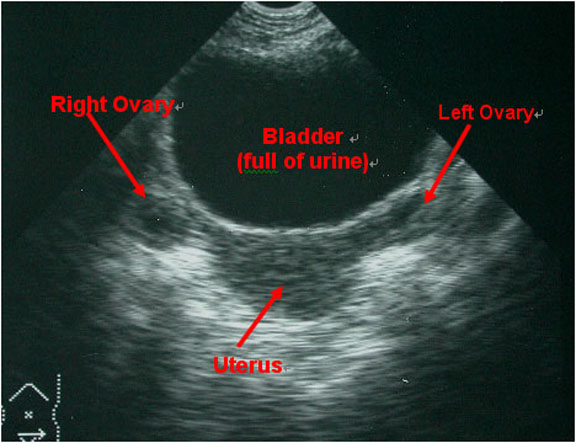
In the absence of masses in the nongravid patient, the uterus, ovaries, and adnexa are situated in the true pelvis. A blunt device called a probe is placed on your skin. It may be used to check for a number of conditions. Abdominal, or transabdominal ultrasounds can produce images of the bladder, uterus, cervix, ovaries and fallopian tubes. How is the pelvic ultrasound performed? These exams are frequently used to evaluate the reproductive and urinary systems. Your doctor is easily able to view the uterus, cervix, vagina, fallopian tubes and ovaries during a pelvic ultrasound. Imaging tests can identify abnormalities and help doctors diagnose conditions. Pelvic pain may be related to many organs. Cpt 76705, under diagnostic ultrasound procedures of the abdomen and retroperitoneum. Ultrasound imaging uses soundwaves to create pictures of the inside of the body. Common causes of pelvic pain. In the pelvic cavity, there are portions of the intestines, the organs of the reproductive, the urinary, and a large part of the musculoskeletal systems.
The true pelvis is bounded anteriorly by the pubis and pubic rami, posteriorly by the sacrum and coccyx, laterally by the fused ilium and ischium, and inferiorly by the muscles of the pelvic floor. And the use of ultrasound to diagnose pelvic pain in women. In women, a pelvic ultrasound may be used for a number of reasons, including those listed below. Your doctor is easily able to view the uterus, cervix, vagina, fallopian tubes and ovaries during a pelvic ultrasound. These exams are frequently used to evaluate the reproductive and urinary systems.
Mobile Pelvic Ultrasound from www.sbadandrmc.com A pelvic ultrasound, also known as pelvic ultrasonography, a pelvic scan or abdominal ultrasound, is a safe and painless diagnostic imaging test used to evaluate the pelvic area for any abnormalities. Common causes of pelvic pain. Ultrasound imaging uses soundwaves to create pictures of the inside of the body. Pelvic ultrasound can help to diagnose a variety of conditions in both men, women, and children. In the pelvic cavity, there are portions of the intestines, the organs of the reproductive, the urinary, and a large part of the musculoskeletal systems. In general for pocus exams, it is usually good to start with the transabdominal ultrasound and then use the transvaginal approach if needed. Transabdominal pelvic ultrasound can detect most larger abnormalities such as large fibroids, ovarian cysts, neoplasms, etc. It is used to examine organs in the pelvis.
Pelvic pain may be related to many organs.
It is used to examine organs in the pelvis. The abdominal, the pelvic/gynaecology and the urinary ultrasound scan. A pelvic ultrasound is a noninvasive diagnostic exam that produces images that are used to assess organs and structures within the female pelvis. 1.) the first part is to look at the pelvic organs from the outside of your abdomen called transabdominal pelvic ultrasound. This ultrasound examination will examine all the abdominal organs visible on ultrasound scanning. Transabdominally (through the abdomen) and transvaginally (through the vaginal canal). Shop our carefully curated collection of high quality products today! Liver, gallbladder, kidneys, bladder, uterus, ovaries, prostate and seminal vesicles. A renal ultrasound is used to evaluate the kidneys, ureters and bladder. Pelvic ultrasound can help to diagnose a variety of conditions in both men, women, and children. Please follow these simple instructions so that we may better serve you. An abdominal ultrasound is performed to evaluate abdominal structures, including the abdominal aorta. However, its views may be limited by abdominal structures such as bowel gas.
Your doctor might order this test to diagnose a condition, or to check the health of your baby. An abdominal ultrasound is used to evaluate the organs and blood vessels in the abdomen, including the gallbladder, kidneys, liver, pancreas and spleen. The sound waves create a picture on a video monitor. Imaging tests can identify abnormalities and help doctors diagnose conditions. Pelvic ultrasound can help your doctor identify problems in your lower abdominal and pelvic organs, such as your bladder.
Female Pelvis Us Toronto Notes from torontonotes.ca However, its views may be limited by abdominal structures such as bowel gas. Both women and men can get pelvic ultrasounds, but it's more common for women to get them. Cpt 76705, under diagnostic ultrasound procedures of the abdomen and retroperitoneum. There are three types of pelvic ultrasound: Liver, gallbladder, kidneys, bladder, uterus, ovaries, prostate and seminal vesicles. Abdominal, vaginal (for women), and rectal (for men). An ultrasound, also named sonography, of the abdomen and the pelvic makes it possible to see your abdominal and pelvic organs: An abdominal ultrasound is used to evaluate the organs and blood vessels in the abdomen, including the gallbladder, kidneys, liver, pancreas and spleen.
Imaging tests can identify abnormalities and help doctors diagnose conditions.
Both women and men can get pelvic ultrasounds, but it's more common for women to get them. Transabdominally (through the abdomen) and transvaginally (through the vaginal canal). A pelvic ultrasound is a test that uses sound waves to make pictures of the organs inside your pelvis. A pelvic ultrasound is a test that uses sound waves to make a picture of the inside of the lower belly (pelvis). Depending on the patient and the condition being assessed, either one or both of these methods can be used. Cascade has served midwives and other healthcare professionals for over 40 years! A renal ultrasound is used to evaluate the kidneys, ureters and bladder. An abdominal ultrasound is used to evaluate the organs and blood vessels in the abdomen, including the gallbladder, kidneys, liver, pancreas and spleen. And the use of ultrasound to diagnose pelvic pain in women. The approach your doctor recommends for your ultrasound depends on the reason for your test and whether you are a man or a woman. Provides a brief description about obtaining images of the uterus and ovaries via trans abdominal ultrasound. A blunt device called a probe is placed on your skin. Shop our carefully curated collection of high quality products today!
An ultrasound machine produces a picture of your body using sound waves pelvic ultrasound female. A blunt device called a probe is placed on your skin.